Digitalização e impressão 3D para a reconstrução das perdas volumétricas num modelo anatómico de cera do século XVIII
DOI:
https://doi.org/10.14568/cp2018003Palavras-chave:
Fotogrametria, Tecnologias digitais, Conservação e restauro, Ceroplástica, Colecções científicasResumo
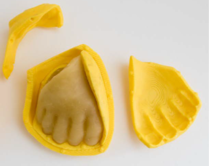
Os modelos tridimensionais de cera pertencentes ao património científico da Universidade Complutense de Madrid são objectos raros e extraordinários que merecem ser preservados porque têm grande relevância para se entender como cirurgiões e anatomistas encontraram fórmulas de construção visual que possibilitaram a difusão do seu conhecimento sobre o corpo humano. Na presente pesquisa, num modelo anatómico de cera do século XVIII, com um notável grau de deterioração e fragilidade estrutural, foi testada uma metodologia de reconstrução volumétrica baseada na digitalização do objecto por fotogrametria, modelagem 3D das peças danificadas e impressão em 3D dos respectivos moldes, para no final se obter cópia em material ceroso. Graças a esta metodologia, foi possível reduzir o manuseamento da escultura durante o processo de restauro e foi minimizado o risco de deterioração acidental.
Recebido: 2018-1-29
Revisto: 2018-4-17
Aceite: 2018-6-2
Online: 2018-6-11
Publicação: 2019-1-10
Downloads
Referências
[1] De Chandarevian, S.; Hopwood, N. (eds.), Models: The Third Dimension of Science, Stanford University Press, Stanford (2004).
[2] Maerker, A., 'Anatomizing the trade: designing and marketing anatomical models as medical technologies, ca. 1700-1900', Technology and Culture 54(3) (2013) 531-562, https://doi.org/10.1353/tech.2013.0108.
[3] Haviland, T. N.; Parish, L. C., 'A brief account of the use of wax models in the study of Medicine', Journal of the History of Medicine and Allied Sciences 25(1) (1970) 52-75, https://dx.doi.org/10.1093/jhmas/XXV.1.52.
[4] Talairach-Vielmas, L., 'Anatomical models: a history of disappearance?', Histoire, Médecine et Santé 5 (2014) 9-20.
[5] 'The Leiden declaration on human anatomy/anatomical collections' (2012), International Conference on 'Cultures of Anatomical Collections', held at Leiden University, Leiden University, http://media.leidenuniv.nl/legacy/leiden-declaration.pdf (acceso en 2018-6-10).
[6] Pierdicca, R.; Frontoni, E.; Malinverni, E. S.; Colosi, F.; Orazi, R., 'Virtual reconstruction of archaeological heritage using a combination of photogrammetric techniques: Huaca Arco Iris, Chan Chan, Peru', Digital Applications in Archaeology and Cultural Heritage 3(3) (2016) 80-90, https://doi.org/10.1016/j.daach.2016.06.002.
[7] Scopigno, R.; Cignoni, P.; Pietroni, N.; Callieri, M.; Dellepiane, M., 'Digital fabrication techniques for cultural heritage: a survey', Computer Graphics Forum 36(1) (2017) 6-21, https://doi.org/10.1111/cgf.12781.
[8] Di Paola F., 'Digital technologies for virtual recomposition: the case study of Serpotta stuccoes', Journal of the Malta Chamber of Scientists 3(1) (2015) 63-68, https://doi.org/10.7423/XJENZA.2015.1.09.
[9] Charbonnier, B.; Laurent, C.; Blanc, G.; Valfort, O.; Marchat, D., 'Porous bioceramics produced by impregnation of 3d-printed wax mold: ceramic architectural control and process limitations', Advanced Engineering Materials 18(10) 1728-1737, https://doi.org/10.1002/adem.201600308.
[10] Rodriguez, R. U.; Kemper, N.; Breathwaite, E.; Dutta, S. M.; Hsu, E. L.; Hsu, W. K.; Francis, M. P., 'Demineralized bone matrix fibers formable as general and custom 3D printed mold-based implants for promoting bone regeneration', Biofabrication 8(3) (2016) 035007, https://doi.org/10.1088/1758-5090/8/3/035007.
[11] Arbace, L.; Sonnino, E.; Callieri, M.; Dellepiane, M.; Fabbri, M.; Iaccarino Idelson, A.; Scopigno, R., 'Innovative uses of 3D digital technologies to assist the restoration of a fragmented terracotta statue', Journal of Cultural Heritage 14(4) (2013) 332-345, https://doi.org/10.1016/j.culher.2012.06.008.
[12] Dacome, L., Malleable Anatomies. Models, Makers, and Material Culture in Eighteenth-Century Italy, Oxford University Press, Oxford (2017).
[13] Azzaroli Puccetti, M. L.,'Human anatomy in wax during the Florentine Enlightenment', Italian Journal of Anatomy and Embriology 102(2) (1997) 77-89.
[14] Maerker, A., Models Experts: Wax Anatomies and Enlightenment in Florence and Vienna, 1775-1815, Manchester University Press, Manchester (2011).
[15] Maerker, A., 'Florentine anatomical models and the challenge of medical authority in late-eighteenth-century Vienna', Studies in History and Philosophy of Science 43(3) (2012) 730-40, https://doi.org/10.1016/j.shpsc.2012.02.005.
[16] Usandizaga, M., Historia del Real Colegio de Cirugía de San Carlos de Madrid (1787-1828), Consejo Superior de Investigaciones Científicas, Madrid (1948).
[17] Burke, M. E., The Royal College of San Carlos. Surgery and Spanish Medical Reform in the Late Eighteenth Century, Duke University Press, Durham (1977).
[18] Sánchez, A.; Del Moral, N.; Micó, S., 'Entre la Ciencia y el Arte. Ceroplástica anatómica para el Real Colegio de Cirugía de San Carlos (1786-1805)', Archivo Español de Arte 85(340) (2012) 329-349, https://doi.org/10.3989/aearte.2012.v85.i340.518.
[19] 'Libro de representaciones, respuestas y ordenanzas, y Reales Ordenes con arreglo a la rdenanza de cirugía. Madrid, 1787-1815', manuscrito, Archivo General de la Universidad Complutense, Madrid, BH UCM, Mss 927.
[20] Massey, L., 'Pregnancy and pathology: picturing childbirth in eighteenth-century obstetric atlases', The Art Bulletin 87(1) (2005) 73-91, https://doi.org/10.1080/00043079.2005.10786229.
[21] Smellie, W., A Sett of Anatomical Tables, With Explanations and an Abridgement of the Practice of Midwifery: With a View to Illustrate a Treatise on That Subject, and Collection of Cases, London (1754).
[22] Bonells, J.; Lacaba, I., Curso Completo de Anatomía del Cuerpo Humano, vol. 5, Imprenta de Sancha, Madrid (1800).
[23] Sánchez, A.; Matia, P., 'Modelos plásticos o simulacros de carne. Procedimientos tecnológicos en la creación de esculturas en cera', De Arte 15 (2016) 310-326, https://doi.org/10.18002/da.v0i15.
Downloads
Publicado
Como Citar
Edição
Secção
Licença
O presente trabalho é distribuído nos termos da Licença Creative Commons (CC BY-NC 4.0) que permite a utilização, partilha e reprodução para fins não comerciais e sem modificações, desde que o autor e fonte original sejam citados.
O Copyright permanece com os autores.